Thigh Anatomy Concise Medical Knowledge
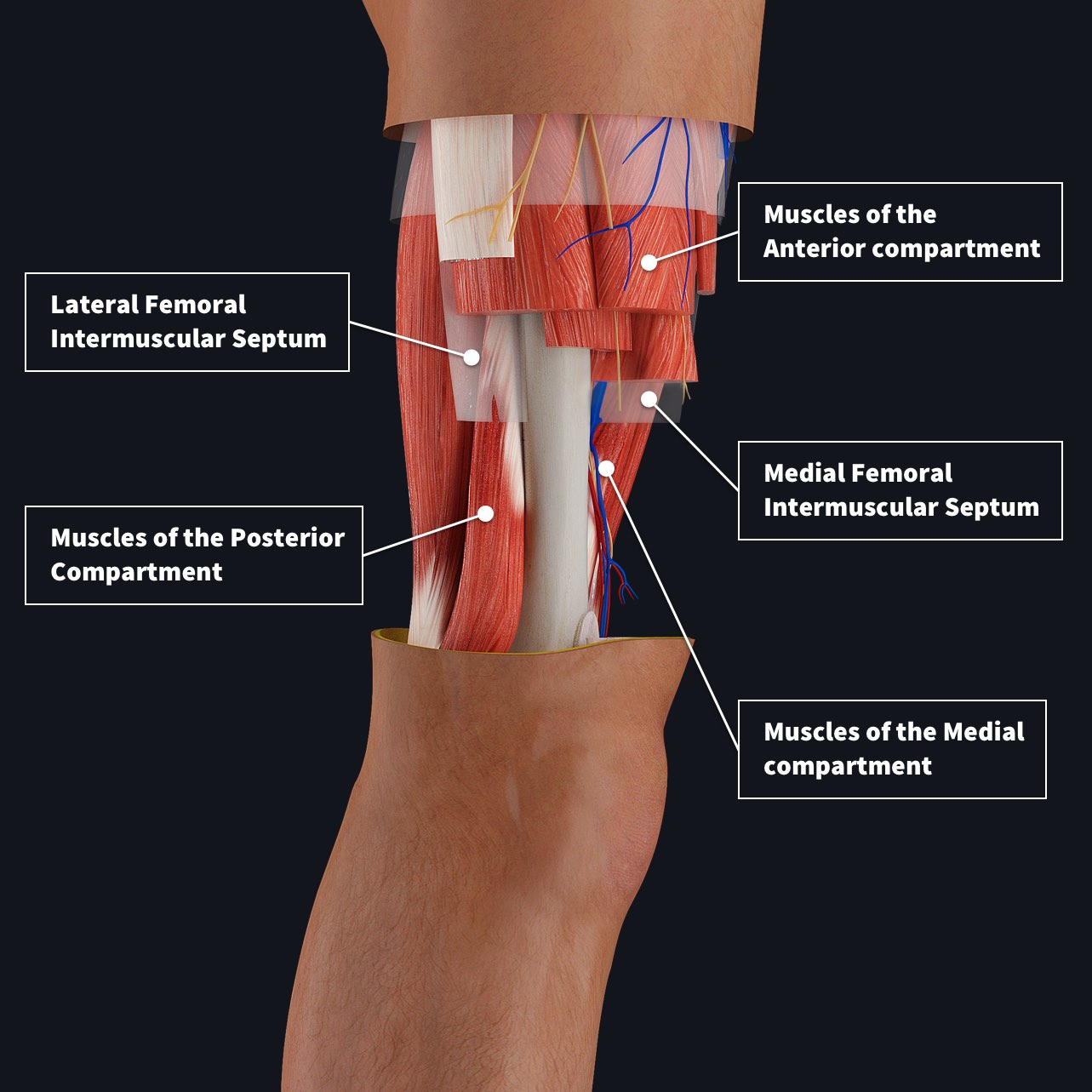
The medial (adductor) compartment of the thigh is one of the three compartments in the thigh. Muscles within this compartment form the adductor group as they primarily produce hip adduction. The thigh is separated into anterior, posterior and medial (adductor) compartments by intermuscular septa and surrounded by the fascia lata. Muscles
medial thigh anatomy
The medial compartment of the thigh consists of the following muscles: Check it out. Previous slide 8 / 22. Muscles of lower limb (overview) Muscles of hip region (part 1) Muscles of hip region (part 2) Muscles of hip region (part 3) Anterior compartment of thigh muscles (part 1).
Muscles of the Medial Thigh TeachMeAnatomy
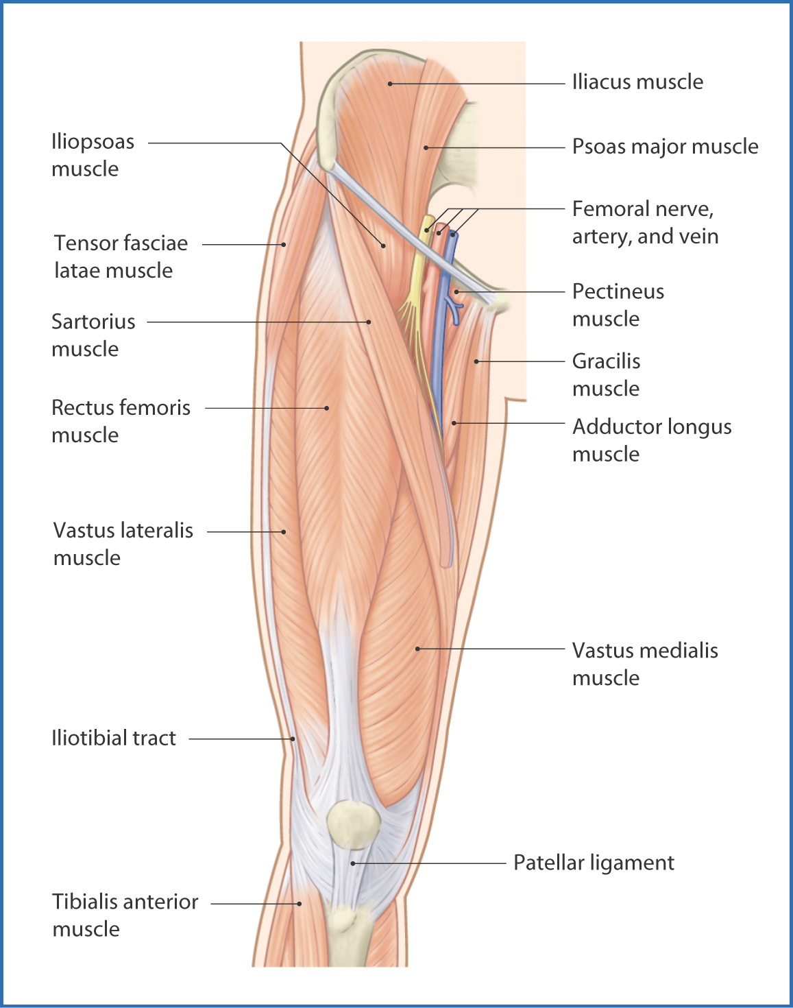
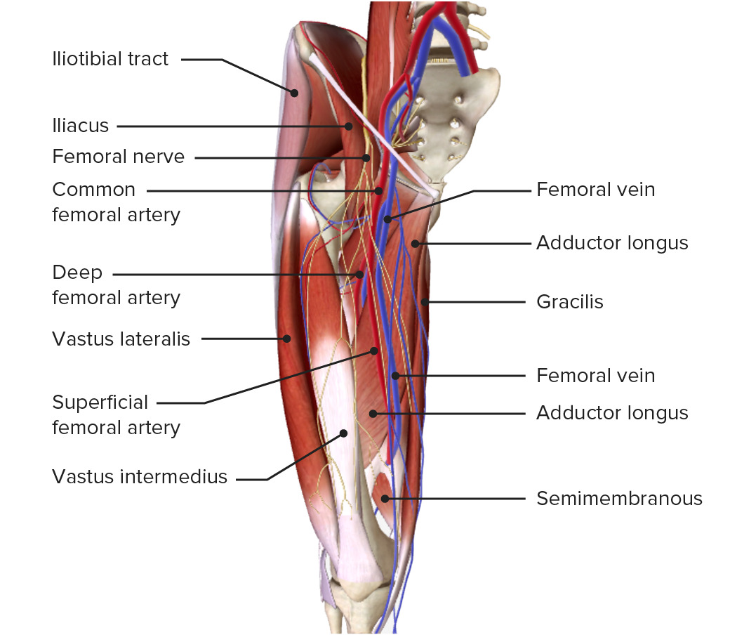
The function of the anterior compartment muscles is to extend the lower limb at the knee joint. The innervation of the anterior compartment of the thigh is from the femoral nerve, which originates from spinal roots L2-L4, and blood supply is from the femoral artery and its first branches.

Medial compartment thigh muscles YouTube
1 Images summary Thigh Compartment Syndrome is a devastating lower extremity condition where the osseofascial compartment pressure rises to a level that decreases perfusion to the thigh and may lead to irreversible muscle and neurovascular damage.

Muscles of the Anterior Thigh Quadriceps TeachMeAnatomy
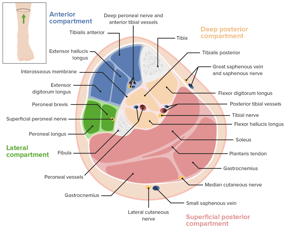
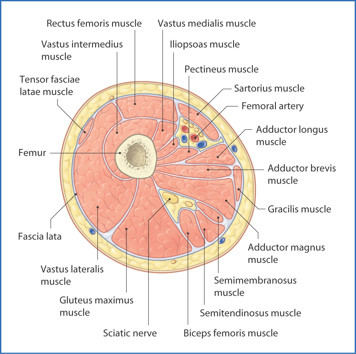
The thigh is the part of the lower limb located between the hip and the knee, and it can be divided into anterior, medial and posterior compartments that surround the femur.These compartments are formed by the intermuscular septa that originate on the inner surface of the fascia lata and attach to the linea aspera of the femur.. And, more importantly, each compartment contains its own muscles.
Muscles of the Medial Thigh TeachMeAnatomy
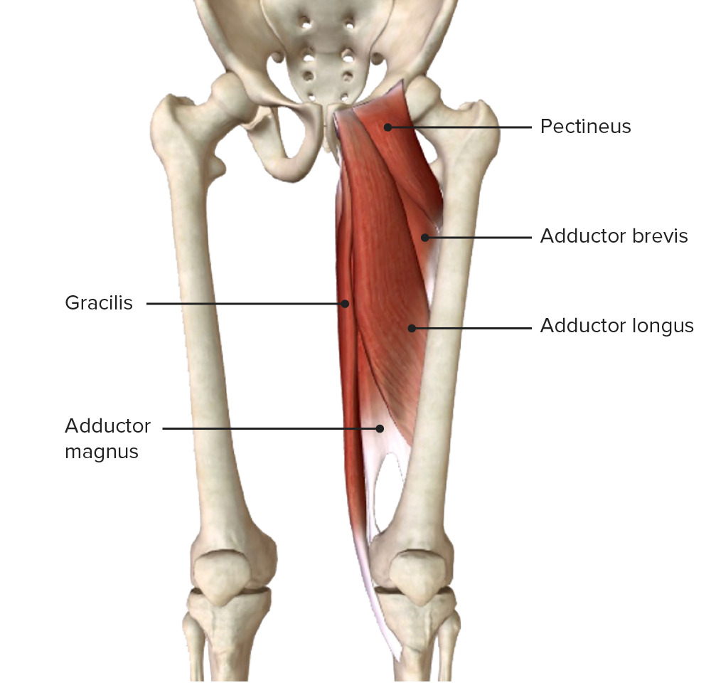
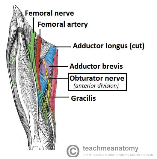
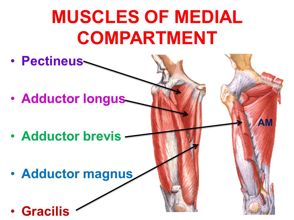
The muscles in the medial compartment of the thigh are collectively known as the hip adductors. There are five muscles in this group; gracilis, obturator externus, adductor brevis, adductor longus and adductor magnus. All the medial thigh muscles are innervated by the obturator nerve, which arises from the lumbar plexus.
Leg Anatomy Concise Medical Knowledge
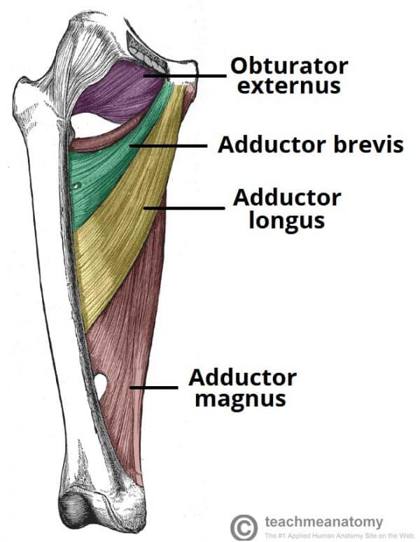
The medial fascial compartment of thigh contains the hip adductors. The obturator nerve is the primary nerve supplying this compartment. The muscles in the compartment are: gracilis. adductors. adductor longus. adductor brevis. adductor magnus. The obturator externus muscle is sometimes considered part of this group,and sometimes excluded.
medialthighadductorcanal
The medial compartment of thigh is one of the fascial compartments of the thigh and contains the hip adductor muscles and the gracilis muscle . The obturator nerve is the primary nerve supplying this compartment. The obturator artery is the blood supply to the medial thigh. The muscles in the compartment are: gracilis adductor longus

Medial Compartment of Thigh Muscles attachments, action and nerve
Muscles in the Medial Compartment of the Thigh. View Article. Muscles in the Posterior Compartment of the Thigh. View Article. Anatomy Video Lectures. START NOW FOR FREE. TeachMe Anatomy. Part of the TeachMe Series.

Muscles of the Medial Thigh TeachMeAnatomy
The thigh has some of the largest muscles in the human body. The medial thigh muscles are essential for normal gait and lower extremity functioning. The medial thigh muscles mainly allow for adduction of the leg. Weak adductor muscles can create knee instability and increase the risk of an adductor strain.[1] The medial thigh muscles also protect important neurovascular structures as they pass.
Thigh Anatomy Concise Medical Knowledge
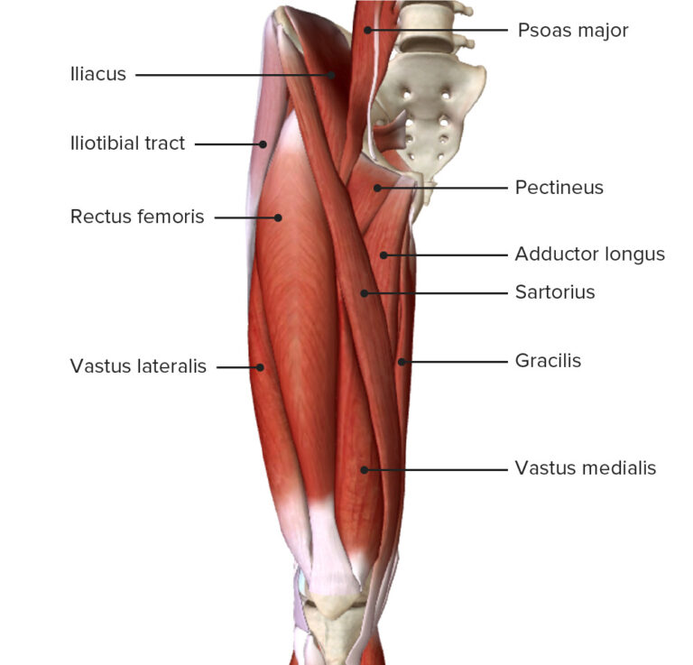
The muscles of the medial compartment (adductor compartment) are one of three subgroups of the thigh muscles, the other two being the muscles of the anterior and posterior compartments. Overall, these muscles mainly act to adduct the thigh at the hip joint. The muscles of the medial compartment of thigh consist of the: - obturator externus.
Thigh Anatomy Concise Medical Knowledge
Green is the medial compartment (gracilis and adductor magnus), blue is the posterior (semimembrosus to biceps c. brevis) and red is the anterior (vastus lateralis to sartorius). The fascial compartments of thigh are the three fascial compartments that divide and contain the thigh muscles.
Medial compartment of thigh muscles, Growth & regeneration of smooth
Anatomically, the leg is defined as the region of the lower limb below the knee. It consists of a posterior, anterior and lateral compartment. In accordance, the muscles of the leg are organized into three groups:
Anteromedial Thigh Basicmedical Key
The hip adductors are a group of five muscles located in the medial compartment of the thigh. These muscles are the adductor longus, adductor brevis, adductor magnus, gracilis, and pectineus . Due to their position, the hip adductors shape the surface anatomy of the medial thigh.
Muscle compartments of the Thigh Complete Anatomy
Mnemonic Sources Related articles + Show all Pelvis The bony framework of the pelvis, called the pelvic girdle, is comprised of two hip bones, the sacrum and the coccyx. The hip bone is made by fusion of three bones; ilium, ischium and pubis. The hip bones articulate with themselves via pubic symphysis, and with the sacrum via the sacroiliac joint.

Medial compartment of thigh Medical anatomy, Muscle anatomy, Thigh
#thigh #anatomy #adductorLink for Donations https://paypal.me/studentlamedicina?locale.x=en_UShttps://www.instagram.com/anatomy.knowledge/The medial compartm.
